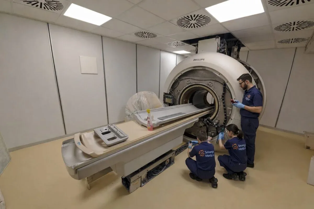
MRI systems and planning
Which MRI scanner should you choose? Low field, 1.5T, 3T, exams and budget
The best MRI system is not always the one with the highest field. A sound choice connects anatomical coverage, image requirements, volumes, gradients, patient access, local competition, site costs and authorisation.
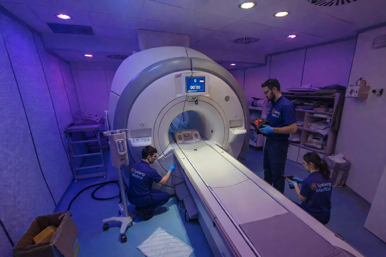
When an organisation assesses an MRI scanner, the first temptation is to rank systems by tesla: low field, 1.5T and 3T. That is rather like choosing a car by looking only at engine power. Static field strength matters, but it does not tell you on its own what the platform can do, how difficult it will be to install, how many patients it can examine or whether the investment makes sense in the facility’s actual market.
The useful question is therefore not “which MRI scanner is the most powerful?” but “which platform can reliably deliver the examinations we intend to provide, at the expected volume and at a sustainable cost?” Answering it requires at least four perspectives:
- the anatomical ROI, or regions of interest and clinical applications the service needs to cover;
- financial return on investment, which uses the same abbreviation;
- local competition and unmet demand;
- the complete project: scanner, site, utilities, authorisation, staff, service and operational continuity.
A dedicated extremity scanner may be an excellent choice for an orthopaedic centre and the wrong one for a provider planning neuro, abdominal and pelvic MRI. A 3T system can create a genuine advantage in a specialist centre, but it may be oversized when the case mix is almost entirely routine and the extra signal is not converted into protocols, expertise or productivity. A modern 0.55T whole-body system is not simply a new version of an older low-field scanner either: it may address different installation, accessibility and application goals.
The short answer: which field for which project?
| Indicative type | Where it may make sense | What needs careful verification |
|---|---|---|
| Dedicated or sector-specific MRI up to 0.5T | Volumes concentrated on extremities and joints supported by the intended use; orthopaedic or physiatry setting; limited space and investment | Actual anatomical coverage, image performance for the intended exams, scan times, positioning, exclusions and correct regulatory classification |
| Open low- or mid-field MRI | Accessibility, specific positioning needs, selected patients, MSK and certain routine exams according to platform | Real anatomical coverage, coils, protocol duration, uniformity, advanced techniques and the distinction between a true open magnet and a wide bore |
| Modern 0.55T whole-body MRI | Projects sensitive to footprint, cryogens, infrastructure or accessibility; an exam portfolio validated for that platform | Do not generalise results from selected applications to every examination; verify software, coils and performance against the planned mix |
| 1.5T whole-body MRI | General diagnostic imaging, broad case mix, established protocols and a balance of performance, robustness and cost | System generation, gradients, coils, licences and workflow: two 1.5T scanners may differ substantially |
| 3T whole-body MRI | Neuro, high-detail MSK, prostate, research and other specialist applications where the additional signal is used effectively | Susceptibility, B0/B1 uniformity, SAR, artefacts, expertise, cost, protocol complexity and proven local demand |
This is an initial map, not a ranking. “Low”, “mid” and “high field” do not have universal thresholds in every context, as an ACR technical publication also notes. It is more informative to state the actual strength — 0.31T, 0.4T, 0.55T, 1.5T or 3T, for example — together with system geometry and intended use. Italian regulatory thresholds are also designed for safety and authorisation; they are not a score for image quality.
What really changes as field strength increases
Tesla describes the strength of the static magnetic field, known as B0. A stronger B0 generally provides more signal. That margin can be used in different ways:
- smaller voxels and higher spatial resolution;
- shorter acquisition time for a sequence;
- greater acceleration;
- support for signal-intensive techniques;
- a different balance between detail, coverage and duration.
It does not mean that twice the field produces images that are “twice as good” or automatically halves the examination time. The real benefit depends on anatomy, sequence, coil, body habitus, movement, reconstruction and how the protocol spends the available signal.
Higher field also introduces challenges. At 3T, susceptibility and chemical-shift effects become more prominent; B0 and B1 uniformity, fat suppression, dielectric effects and radiofrequency deposition limits can require more optimisation. In certain anatomical areas or near metal, gas and air–tissue interfaces, a lower field may be more tolerant. Scientific reviews comparing 1.5T and 3T describe this trade-off between signal reserve and technical complexity, rather than a universal winner.
From a safety perspective, “lower field” does not automatically mean “safe with every implant”. An MR Conditional device must be identified exactly and used within all conditions stated by its manufacturer. These may include static field strength, gradients, RF limits, position and protocol. The FDA overview of MRI benefits and risks and the ACR Manual on MR Safety are useful international references.
Low field is not one type of scanner
Very different systems are grouped under the “low-field” label: permanent extremity scanners, true open magnets, weight-bearing systems and newer whole-body platforms. Their strengths and limitations should not be treated as interchangeable.
Potential advantages include:
- permanent magnets with no cryogenic helium in some platforms;
- favourable access and positioning with certain geometries;
- potentially lower footprint, power and infrastructure requirements;
- reduced susceptibility and off-resonance effects;
- lower RF energy deposition and potential benefits in selected applications;
- a strong economic model when the examination portfolio is tightly focused.
The central physical constraint is lower signal-to-noise ratio. Depending on the scanner and protocol, it may require more time, larger voxels or a narrower range of advanced applications. Modern coils, gradients, optimised sequences and reconstruction have improved low-field performance substantially, but they do not make every platform equivalent to 1.5T or 3T for every anatomy.
A dedicated scanner for knee, ankle, hand, wrist and elbow may be very productive in an orthopaedic service with the right case mix. Shoulder, hip, spine, patients with restricted mobility and anything beyond its field of view or intended use must be checked separately. A lower-priced scanner that cannot perform a material share of requested exams is no longer inexpensive: it sends patients elsewhere and reduces the service’s value.
“Open” also needs precision. A true open magnet, a short- or wide-bore cylinder, a dedicated extremity scanner and an upright system are different designs. Bore diameter and length, remaining space with the coil, table limit, centring and patient position may matter more than the marketing label. Wider access can help many people, but it does not always remove claustrophobia, pain or the difficulty of remaining still.
Why 1.5T remains the general-purpose choice
For many imaging providers, 1.5T whole-body MRI remains the natural balance. It supports established protocols across a broad examination range, offers extensive coil and application options, draws on widespread operator experience and has a substantial new and refurbished equipment ecosystem.
With the right system, accessories, software and expertise, it can support routine neuro, spine, large and small joints, abdomen, pelvis, breast, vascular and cardiac work. In the presence of prostheses, gas or difficult anatomy, 1.5T may also be more predictable than 3T for certain sequences.
“1.5T” is not a configuration, however. A platform with limited gradients, few coils, old software and uncertain parts availability is not equivalent to a more recent scanner with modern RF chains, acceleration, advanced applications and structured support. A refurbished-system assessment should identify at least:
- year, software release and upgrade path;
- gradients and the conditions under which their performance is stated;
- included coils, channels, condition and anatomical coverage;
- licences for diffusion, cardiac, breast, angiography, spectroscopy and other required applications;
- magnet configuration, cryogenic history and cold head where fitted;
- table, chiller, compressor and subsystem condition;
- documentation, deinstallation, transport, reinstallation and acceptance testing;
- service and spare-parts availability over the investment horizon.
A well-configured and well-supported 1.5T can therefore deliver more useful examinations and fewer repeats than a 3T selected only for the field-strength label.
When 3T creates a genuine advantage
3T MRI provides a larger signal reserve. It can be used for finer detail, acceleration or demanding techniques and may be particularly valuable in neuroimaging, MR angiography, advanced diffusion, spectroscopy, prostate imaging, small MSK structures and selected cardiac, breast or research applications.
It makes particular sense when the centre has all of the following:
- sufficient demand for specialist examinations;
- radiologists and radiographers able to develop and maintain optimised protocols;
- a referral pathway that values that expertise;
- coils and software packages aligned with the promised applications;
- enough time and organisation to handle complex exams without blocking routine work;
- a financial model capable of supporting the additional investment.
3T is not a marketing shortcut. Extra signal may be spent on resolution rather than time, while SAR, shimming, artefacts and more involved sequences may reduce the throughput benefit. For prostate MRI, for example, the ACR technical parameter recognises that both 1.5T and 3T can produce consistently high-quality examinations when the platform and protocol are appropriate. Field strength alone does not settle the decision.
Before buying 3T, ask which examinations will actually be different, how many will be performed each week, who will report them, what clinical or financial value they create and what competitors already provide. If several underused 3T systems already operate locally but patients still lack access, short waiting times or focused MSK expertise, the competitive opportunity may lie elsewhere.
The first ROI is anatomical; the second is financial
In MRI, ROI normally means region of interest. In an investment plan, it also means return on investment. These two meanings need to be studied together.
Which regions and examinations will you offer?
| Area | Questions to answer before selecting a scanner |
|---|---|
| Extremities and MSK | Small or large joints? Shoulder and hip? Post-operative metal? Weight-bearing exams? Enough volume for a dedicated system? |
| Spine | Routine, post-operative, cord, diffusion or advanced applications? How important is tolerance of hardware artefact? |
| Neuro | Routine brain and spine only, or SWI, perfusion, spectroscopy, fMRI and advanced diffusion? |
| Abdomen and liver | Are diffusion, dynamic contrast, robust fat suppression and short breath-holds required? |
| Pelvis and prostate | What quality is needed, which coils and protocols are available and how important are prostheses or gas? |
| Breast | Are a dedicated coil, injector, dynamic imaging, biopsy and reporting workflow included? |
| Cardiac and vascular | Does the service have expertise, gating, monitoring, injector, software and adequate room time? |
| Lung or regions near air/metal | Does the specific platform provide validated sequences that exploit lower-field and reduced-susceptibility behaviour? |
These are technology-planning questions, not clinical recommendations for individual patients. The appropriate modality and protocol remain the responsibility of qualified healthcare professionals.
How many examinations can you actually complete?
Patients per day cannot be calculated by adding brochure acquisition times. Real throughput includes:
- registration, history and safety screening;
- preparation, IV access and contrast where required;
- changing, connecting and positioning coils;
- moving, centring and removing the patient;
- shimming and protocol-specific preparation;
- repeated sequences caused by movement or artefacts;
- cleaning, reconstruction and image transfer;
- sedation or additional assistance where applicable;
- downtime for maintenance, faults and upgrades.
The useful comparison is a demonstration with representative patients and complete protocols, measuring table time, room occupancy, repeats and accepted image quality. A 3T may be faster for one sequence while being assigned more complex cases. A low-field scanner may acquire more slowly but achieve simple turnover in a highly standardised case mix.
Local competition changes the right answer
Technology must be read within its territory. Before selecting a scanner, map:
- field strengths and geometries offered by competitors;
- examinations with long waiting lists;
- patients currently referred out of the area;
- specialties generating demand: orthopaedics, neurology, oncology, urology and cardiology;
- services that are actually prescribed and reimbursed;
- unresolved access or comfort constraints;
- the clinical reputation the centre can build, not merely advertise.
In an area without accessible MSK services, an open or dedicated platform may address a genuine need. In a neuroscience hub, a correctly equipped 3T may establish a specialist service line. In a high-turnover general imaging centre, a robust, fully equipped and well-supported 1.5T may be the most productive investment.
What gradient specifications really mean
Gradients are time-varying magnetic fields used to localise signal and form the image. They are distinct from the static-field tesla value and can make two scanners carrying the same “1.5T” or “3T” label behave very differently.
The two most advertised specifications are:
- gradient strength, in mT/m: the maximum available amplitude;
- slew rate, in T/m/s: how quickly the gradient can change.
Greater amplitude may help diffusion and spatial encoding. A higher slew rate can support fast sequences such as EPI and gradient echo and may reduce rise time, TE or echo spacing under specific conditions. The real benefit also depends on whether strength and slew are available simultaneously, as well as linearity, usable field of view, duty cycle, cooling, eddy currents, acoustic noise and peripheral-nerve stimulation limits.
A brochure number is therefore insufficient. Ask:
- whether the value is per physical axis or an “effective” vector value;
- what amplitude remains available at the maximum slew rate;
- the rise time for the proposed configuration;
- how long the system sustains demanding sequences before thermal derating;
- linearity and coverage across the required field of view;
- achieved times on the protocols the centre will actually run.
In short, higher-performance gradients can widen protocol options, but do not guarantee more examinations per hour on their own. Siemens’ technical paper on gradient performance and amplifier power likewise stresses the need to consider strength, slew, rise time and operating conditions together.
Coils, software and workflow matter as much as the magnet
The receive coil is where the scanner obtains signal from the patient. Element count and layout, anatomical coverage, proximity to the region of interest, comfort and acceleration capability directly affect image quality and daily work.
An attractively priced scanner may offer poor value if the breast coil is missing, the shoulder coil is faulty, the spine configuration does not cover the required region or changing anatomy requires slow, heavy handling. With refurbished systems, coils should be identified individually, tested and confirmed as supported by the included software release.
The same is true of licences. “Cardiac”, “diffusion”, “angiography”, “metal artefact reduction” and “prostate” do not necessarily describe an included package. Separate what the product family was capable of supporting from what is installed and transferable on the specific unit.
Console design, automation, reconstruction, RIS/PACS integration, stored protocols, training and service quality also matter. The most sophisticated scanner on paper may yield a poorer return if operators cannot exploit it or a critical spare part takes weeks to source.
The real budget is the project cost, not the magnet price
The purchase price is only one part of the required capital. A fair comparison needs a total cost of ownership based on the actual site.
| Cost area | What it may include |
|---|---|
| MRI system | Magnet, gradients, RF, table, console, workstation, coils, licences, accessories and injector |
| Logistics | Deinstallation, packing, transport, insurance, lifting, façade or roof opening and internal moving |
| MRI room | RF shielding or Faraday cage, finishes, doors, observation window, lighting, floor and access control |
| Stray field | Field-line planning and active or passive magnetic shielding where required |
| Cryogenics | Quench pipe where required, sensors, ventilation, helium, cold head, compressor and emergency arrangements according to technology |
| Utilities | Electrical supply, panels, UPS where specified, earthing, HVAC, chiller and process water where necessary |
| IT and integration | Network, cybersecurity, DICOM, RIS/PACS, storage, worklist, reporting and backup |
| Design and authorisation | Architectural and engineering design, appointed experts, applications, verification and documentation |
| Commissioning | Installation, shimming, acceptance, quality controls, training and protocol development |
| Operations | Energy, water, maintenance, on-call support, parts, helium, downtime, upgrades and insurance |
| End of life | Deinstallation, magnet management, transport, site reinstatement and residual value |
Not every item applies in the same way to every scanner. A permanent magnet may need no cryogenic installation; a sealed low-helium design may reduce or change traditional requirements; some systems depend on external chillers while others use different cooling solutions. Only the manufacturer’s site planning and the appointed professionals can define what the individual project requires.
The budget should also include revenue lost during downtime. A more expensive service agreement may be economically sound if it reduces response times and operational risk. Likewise, a low-priced used scanner remains a good investment only if its configuration, parts, software and support remain available for the planned service life.
Helium-free, low-helium and zero boil-off are not synonyms
Helium-free is used for different technologies and should be read carefully.
- A permanent low-field magnet may operate genuinely without cryogenic helium.
- A conventional superconducting zero-boil-off system normally retains a substantial helium inventory and uses a cold head to recondense it. It reduces routine evaporation but is neither helium-free nor cryogen-free.
- Some recent superconducting platforms use a sealed circuit with a very small helium inventory and are marketed for “helium-free operations”. Philips states approximately 7 litres for its BlueSeal magnet; Siemens describes less than one litre in MAGNETOM Free.Max. That does not mean there is no helium inside the magnet.
New sealed low-helium platforms may carry a higher acquisition price than older conventional or refurbished systems. This is not a universal rule, and the appropriate comparison is the installed and lifecycle cost: helium availability, quench pipe, space, building works, recovery after a loss, energy, maintenance and the value of reduced operational risk may offset part of the initial premium.
Ask the supplier about:
- the actual helium quantity and circuit design;
- behaviour after power loss or cooling failure;
- ramp-down, quench and recovery procedures;
- quench-pipe and ventilation requirements;
- skills, time and cost required to return to service;
- warranty and responsibility for cryogenic performance.
You can explore the operational topics further in our articles on MRI cold-head maintenance, helium refill and ramp-up and ramp-down.
Dedicated MSK or whole body: clinical and Italian regulatory differences
A scanner may be marketed as “MSK”, “dedicated” or “open”, but the legal category used by the Italian Ministerial Decree of 14 January 2021 is sector-specific MRI equipment. The decree associates it with imaging limbs, shoulders and hips, and spinal biomechanics in supine and standing positions.
The authorisation exemption under Article 2 applies only when the stated conditions are met together, including:
- a new-generation sector-specific system;
- static field not exceeding 0.5T;
- a non-superconducting magnet;
- intended use for the examinations listed in the decree.
The exemption does not remove applicable safety standards, professional responsibilities or other required steps. A whole-body scanner used mostly for knees and shoulders normally remains non-sector-specific: the local examination mix does not by itself change the equipment classification.
For non-sector-specific MRI up to 4T, the national rule provides for installation at an authorised healthcare facility also equipped with CT, conventional radiography and ultrasound. The Region may regulate an exemption from the CT-presence requirement through integration with nearby imaging facilities; this is not automatic and needs project-specific confirmation.
This distinction may radically alter the business plan, rooms and authorisation timetable. The official classification should therefore be confirmed before budget is committed, rather than inferred from a commercial description. Our article CT or MRI? Differences, uses and Italian requirements discusses the requirement in more detail.
Model examples, not a ranking
The following names illustrate how different MRI platforms can be. They are not a complete comparison, recommendation, stock statement or offer. Configuration, licences, specifications, commercial status and availability in Italy must be checked for the individual unit and with the manufacturer.
| Field and configuration | Example families/models | What they illustrate |
|---|---|---|
| Dedicated extremity, 0.31T | Esaote O-scan | Permanent magnets without cryogens and an extremity-focused workflow, with coverage unlike a whole-body system |
| Permanent open, 0.4T | Fujifilm APERTO Lucent | True open geometry and no cryogenic helium in a low-field platform |
| Modern whole body, 0.55T | Siemens MAGNETOM Free.Max | A newer interpretation of lower field, wide bore and a sealed magnet with minimal helium |
| Whole body, 1.5T | Siemens MAGNETOM Aera, Philips Ingenia Ambition, Canon Vantage Orian, GE SIGNA Artist | Four examples of general-purpose platforms; generation, gradients, magnet, coils and software remain decisive |
| Whole body, 3T | Siemens MAGNETOM Skyra, Philips Ingenia Elition, GE SIGNA Premier, Canon Vantage Galan 3T | High-field platforms for advanced general or specialist work, with differing configurations and performance |
Official material presents Esaote O-scan as a 0.31T dedicated extremity system, Fujifilm APERTO Lucent as a permanent 0.4T open system and Philips Ingenia Ambition as a 1.5T system using a BlueSeal magnet. Year, release, gradients, coils and packages can still vary within a product family; the commercial name alone does not describe the proposed unit.
Our sourced systems area presents models such as Philips Ingenia 1.5T and Philips Ingenia 3.0T as references for a sourcing request. This does not mean that they are held in stock: availability is searched and qualified against the required configuration.
Four scenarios that illustrate the method
1. Orthopaedic centre with high extremity volume
If demand is concentrated on knee, ankle, foot, hand, wrist and elbow, a dedicated permanent system may offer a strong relationship between space, investment and productivity. The team should first measure how many referrals involve shoulder, hip, spine or unsupported regions and verify the individual scanner’s quality, comfort and regulatory pathway.
2. General diagnostic imaging centre
When neuro, spine, MSK, abdomen and pelvis are spread across the week, a complete 1.5T whole-body system is often the most balanced starting point. Coils, gradients, software, bore, reliability and support are more useful differentiators than field strength alone.
3. Specialist or referral centre
When documented volumes exist in advanced neuro, prostate, high-detail MSK or research, 3T may create a clinical and commercial advantage. A pathway must still turn the available signal into protocols, expertise, referrals and financial sustainability.
4. Site with major building or cryogenic constraints
A permanent or sealed low-helium system may reduce some constraints and risks, but feasibility should not be inferred from a brochure. The project still needs field-line planning, loading and access checks, RF design, HVAC, electrical supply, cooling and an authorisation review. Saving one item of building work does not prove the lowest total cost.
Checklist before requesting a quotation
- List examinations by anatomical region and complexity, not merely “MSK” or “whole body”.
- Estimate credible monthly volumes, peaks, seasonality and three-to-five-year growth.
- Map competitors, waiting lists, referrals and unmet local demand.
- Decide which advanced applications will genuinely create value.
- Compare complete protocols and room time, not isolated demonstration sequences.
- Inventory all included coils, licences, injector, workstation and accessories.
- Check gradients, field of view, bore, table, noise and comfort against the use case.
- Establish Italian sector-specific or non-sector-specific classification and the regional authorisation route.
- Commission a preliminary site design covering access, RF cage, field lines, utilities, chiller/HVAC and cryogenics.
- Calculate capital cost, operating cost, downtime, service agreement and end-of-life cost.
- For refurbished equipment, assess history, magnet, helium, cold head, coils, software, documents and parts support.
- Request a proposal that clearly separates equipment, services, exclusions, responsibilities and acceptance criteria.
Frequently asked questions
Is 3T always better than 1.5T?
No. It offers more potential signal for resolution, acceleration and advanced techniques, but also adds susceptibility, uniformity and RF complexity. A well-configured 1.5T is fully capable for a broad range of routine work and may be more predictable in some applications.
Does low-field MRI produce worse images?
Lower field provides less signal, but quality and fitness for purpose depend on platform, anatomy and protocol. A modern dedicated scanner may be excellent within its scope; it should not be presented as equivalent to a whole-body scanner for exams it does not support.
How many tesla are needed for MSK MRI?
It depends on the anatomical region, required detail, metal and volume. Selected extremities may suit dedicated systems; shoulder, hip, spine and a wider service may require whole-body capability. Coils and gradients are also decisive.
Do stronger gradients mean faster examinations?
They may enable faster or more efficient sequences, but total time also depends on RF, coils, SAR, reconstruction, protocols, preparation and the patient. Headline values must be interpreted under their actual operating conditions.
Does helium-free mean there is no helium?
Only with certain technologies, such as some permanent magnets. In superconducting sealed low-helium systems, the term can describe operation without routine refills even though a small quantity of helium remains sealed inside the magnet.
Must an Italian facility have CT to operate whole-body MRI?
For non-sector-specific MRI up to 4T, the January 2021 decree includes CT, conventional radiography and ultrasound among the standard equipment of the authorised facility. A Region may regulate an exemption for CT alone through integration with a nearby facility. The current regional rules and the individual project must be checked before investment.
Is a new or refurbished scanner better?
It depends on budget, time horizon, configuration and acceptable risk. A properly assessed refurbished system may provide an excellent performance-to-investment ratio; a new platform may offer efficiency, newer cryogenic technology, warranty and upgrade options. The comparison should include all site and operating costs.
From scanner selection to a complete project
Spinergy Medical can support healthcare providers and investors with technical assessment of new or refurbished MRI systems, configuration sourcing through its commercial network, preliminary site checks and the planning of deinstallation, transport, installation and specialist services.
Our work starts from the real requirement: examinations, volumes, location, timescale and budget. Explore technical consulting, MRI installation planning or describe the system you need in a quotation request. If you are facing these decisions, call or email us: we can turn a list of model names into a project-based comparison.
Medical, regulatory and product information notice
Regulatory information: this content is provided solely for general information and was reviewed against the official sources listed, with an editorial update date of 7 September 2026. It is not legal or regulatory advice, does not establish compliance of any particular facility or scanner, does not confirm eligibility for a regional exemption and does not replace any authorisation or decision by the competent authority. Equipment classification and intended use, field strength, magnet technology, fixed or mobile configuration, facility authorisations, regional rules and site-specific decisions must be checked for the individual project against the law then in force. Before purchasing, installing or placing an MRI system into clinical service, the case should be reviewed with appropriately qualified professionals and the authority or authorities identified under the applicable national and regional rules.
Medical information: clinical content describes general planning criteria and does not replace a prescription, medical assessment or validated protocol. The modality, field strength and protocol appropriate for an individual patient must be selected by healthcare professionals according to the clinical question, the person’s circumstances and any implants.
Products and specifications: brands and models are mentioned solely as technology examples. Specifications, configurations, licences, intended uses, availability and commercial status may vary by country, year and individual unit. They must be confirmed against current manufacturer documentation and the proposal for the specific scanner.
Main sources
- Italian Official Gazette — Ministerial Decree of 14 January 2021 and MRI safety standards
- Italian Ministry of Health — high-field MRI equipment
- INAIL — MRI safety
- FDA — MRI benefits and risks
- Scientific review — low-field MRI opportunities and limitations
- Scientific review — clinical MRI at 3T
- Siemens Healthineers — gradient performance and amplifier power
- Esaote — O-scan
- Fujifilm — APERTO Lucent
- Siemens Healthineers — MAGNETOM Free.Max
- Philips — BlueSeal magnet technology